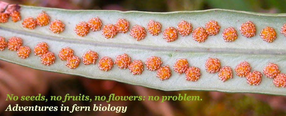

This was an exciting week in the lab! I have two undergraduates working with me this semester, and they just started a project on fern mating systems, the first big project I'm undertaking since coming to UF. The goal is to determine how often ferns undergo an extreme form of selling, called intragametophytic selfing, and whether the tendency to do so has any ecological or phylogenetic correlates. Ferns (and some lycophytes) are the only plants that can do this extreme form of selfing, which leads to homozygosity at all genetic loci (basically, a tremendous loss of variation that leaves them vulnerable to the effects of deleterious alleles).
The first step in the project was to gather a bunch of spores from different fern species, which has been facilitated by many colleagues and collaborators over the last few weeks. Next, the spores are plated onto a nutrient-rich agar medium, and place in a growth chamber, where hopefully they will germinate into gametophytes. The spores that you see in the pictures (the little triangular-shaped structures) are haploid, having developed via meiosis while inside the sporangia (the larger structures that appear to be cracked open in some cases – that's how the spores got out). The sporangia were attached to the leaves of the adult, sporophyte ferns. The gametophytes that develop from these spores will eventually produce eggs and/or sperm that will undergo fertilization, restoring the diploid state and leading to development of the adult sporophyte. That's the fern life cycle in a nutshell!


